Microfluidic platform for perfusable microvascular self-assembly
May 30, 2018|Report
Colette Bichsel1,2, Sean Hall3,4, Ralph Schmid3, Olivier Guenat1,2,3, Thomas Geiser2
1ARTORG Center for Biomedical Engineering Research, University of Bern, Bern, Switzerland
2 Pulmonary Medicine Division, University Hospital of Bern, Bern, Switzerland
3 Thoracic Surgery Division, University Hospital of Bern, Bern, Switzerland
4 Department of Clinical Research, University of Bern, Bern, Switzerland
The project objective is to develop a platform for the formation and maintenance of blood microvessels. The platform is used as physiologically relevant in vitro model of the microvasculature to understand the vascular dynamics in health and disease. The microvessels grown on chip are functional and perfusable from the outside with a defined pressure drop1.
To define the flow channels and chambers for hydrogel and cells, soft lithography is used. Briefly, negative resist (SU-8) is spin-coated on a silicon wafer at 100 μm height and structured using photolithography (Fig.1). The wafer is then used as mold for Poly-dimethylsiloxane (PDMS) that is poured on the wafer, cured, cut and finally bonded to a glass slide (Fig.2).
One of the key issues during the chip production is the complete filling of all the SU8 microstructures with the uncured PDMS. Air bubbles that are often trapped in PDMS must be avoided. For this, the Thinky mixer, which enables to simultaneously mix and degas the PDMS, was used successfully during the process.
The resultant chips are then sterilized and used to culture cells in the defined compartments (chambers of 2 mm diameter and 100 μm height). Endothelial cells and fibroblasts in a fibrin hydrogel are filled in these compartments, and the flow channels are filled with cell culture medium. Within one week, perfusable microvascular structures assemble (Fig.3).1
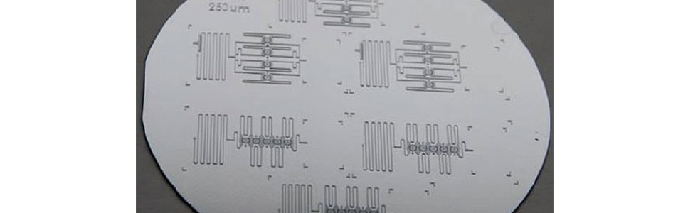
Fig. 1: View of a Si-wafer with SU8 microstructures.
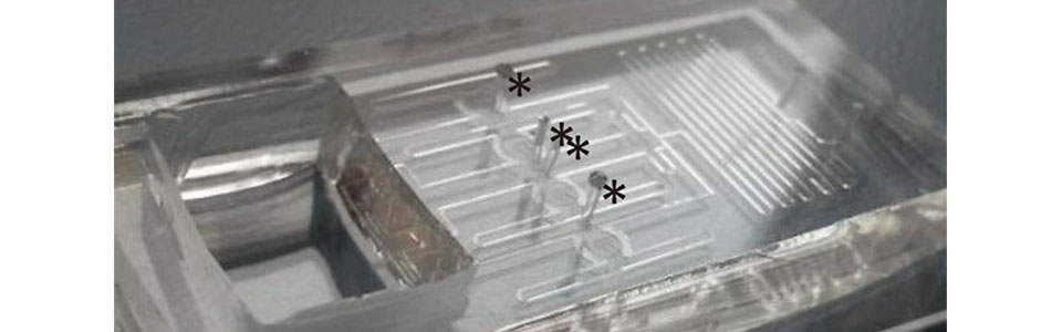
Fig. 2: The vascular chambers are filled with endothelial cells and fibroblasts suspended in a fibrin matrix. Gel inlets are labeled with an asterisk.
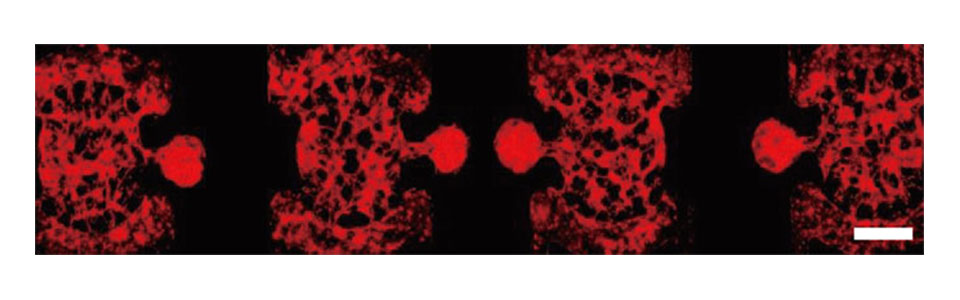
Fig. 3: Rfp-labeled VeraVec endothelial cells self-assemble into 3D microvascular structures. Scale bar: 1 mm